Pathogenesis and treatment of spontaneous quarter cracks - quantifying vertical mobility of the hoof capsule at the heels |
||||||||||||||||||||||
Summary:Spontaneous quarter cracks (Q.C) are defined as cracks, parallel to the horn tubules, originating at the coronary band, in the region of the quarters-heels of the hoof, due to internal trauma as opposed to external trauma (wire cuts e.g.). They consist in a fracture of the hoof wall, from the inside out, proximal to distal, and occur when the ipsilateral ungular cartilage (U.C.), expanding outwards in the loaded limb meets an excessively upwardly shunted hoof wall in the quarter-heel region (Sheared heel) as shown with resected hooves under load. Palpation and measurement of the free margin of the ipislateral, U.C. above the proximal border of the hoof capsule (H.C.) is clinically important in the evaluation and treatment of spontaneous Q.C. Feet with spontaneous Q.C. have a free U.C. margin of less than 15mm depending on the hoof’s size. Increasing the free margin of the U.C. is fundamental in the treatment of spontaneous Q.C. as the lesion originates from the inside out and no amount of crack debridement and fixation by itself will allow for free lateral expansion of the ipsilateral U.C. without it traumatising the coronary band. Lowering of an upwardly shunted heel/quarter can be achieved by special trimming and shoeing methods and /or by a full-wall depth sub-coronary grooving. The first allows for an average of ±7mm of instant hoof wall lowering at the affected heel relative to the ipsilateral U.C. and distal phalanx (D.P.). Finally the author studied the opposite effect; the amount of instant upward displacement of the hoof wall at the heels, relative to the distal phalanx, on the loaded foot with a 20mm heel raise. This was done in vivo by radiological means under standardised trimming and exposure conditions. Average, instant vertical displacement on 18 limbs amounted to 4,3 mm. These results call into question the effectiveness of some raised heel shoeing techniques which aim to change the phalangeal alignment between the D.P. and the proximal phalanges. Abbreviations:
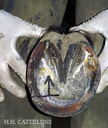
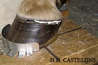
Keywords:Quarter Cracks, Ungular Cartilage, Sheared Heels, Raised Heels, Crack repair, Hoof capsule, Palmar/plantar angle. Quarter Cracks and Hoof shape:Hoof cracks or sand cracks are fractures of the hoof wall, they can be classified; by the region of the hoof which is affected:- toe, quarters, heels or bars; by their direction:- longitudinal (parallel to the horn tubules) or transverse; by their origin:- descending (from the coronary band down) or ascending (from the distal border up); by their depth:- superficial or deep; by their cause:- from external trauma or spontaneous (inside trauma). This paper will only address spontaneous, descending quarter or heel cracks, which, as they originate from the inside out, are always deep. Spontaneous Q.C. are also often bleeding, painful and therefore cause for lameness, while their debridement may show a larger extension close to the laminae then at the surface of the hoof. The incidence of Q.C. is intimately related to hoof shape [O’Grady, 2006] which again is dependent in large part on leg conformation. Hoof shapes adapt to leg conformation in a predictable way and specifically inward or outward rotations of the distal limb will result in hoof shapes that show hyper-development of one heel and it’s opposite toe and underdevelopment of the opposite heel and ipsilateral toe. The underdeveloped diagonal on the bottom of the hoof is parallel to the sagittal axis of the horse, [Castelijns, H., 1999] and this adaptation of the hoof shape to a conformation defect of the limb is useful for the horse as it diminishes excessive distal joint stress. Lateral medial trimming in all horses but especially in horses with angular deviations and/or rotations, should aim at evening out joint spacing in the inter-phalangeal joints of the limb [Caudron, 1998]. Limb conformation defects not only lead to a, (useful), adaptation of the hoof shape to the limb, but also easily deform the hoof capsule, especially when left too long through irregular trimming/shoeing intervals. Deformation (distortion) as opposed to adaptation of the hoof can be defined by bent horn tubules (eg. flares, under-run heels, concave dorsal walls) and vertically distorted coronary bands. [Castelijns, 2006] The later can be objectively recorded by observing and palpating the coronary band as seen from the side, comparing and measuring the bulb’s height as seen from behind. Literature both old and recent has made the connection between sheared heels and Q.C., with clinical experience showing that the crack inevitably originates at the highest point of the coronary band, as seen from the affected side, [Dollar, 1897] [O’Grady, 2006] which perhaps a higher incidence for the medial heel/quarter [Mensa,1950]. (Fig. 1. a, b, c) |
||||||||||||||||||||||
|
||||||||||||||||||||||
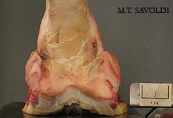
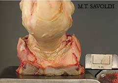
Likewise the observations that the U.C. is involved in the genesis of spontaneous Q.C. isn’t new either; although the author developed this hypothesis on his own, it has been described in literature before [Pires, A., 1991] with references to authors of the later 19 th century, proving the oft repeated truism that “95% of what we know about the art and science of horseshoeing was known by 1900”. [Pieh, W.S., 1993] Ungular Cartilage implications in spontaneous quarter and heel cracks.On loading the limb at the mid-step the fetlock comes down (extends) and the proximal phalanges move down, flexing the distal inter-phalangeal joint. In this stance phase the intermediate phalanx compresses the digital cushion, the distal borders of hoof widen out (hoof mechanism) while the coronary band slightly narrows and the U.C. are pushed outwards. [Barone, R., 1995], [Dollar, A.W., 1897], [Thomason, J.J., 1998] In normal hooves there is ample space above the coronary band for the U.C. to move outwards. This movement has been elegantly recorded on dissected limbs by M.T Savoldi. [Savoldi, 2005] (Fig. 2. a, b) |
||||||||||||||||||||||
|
||||||||||||||||||||||
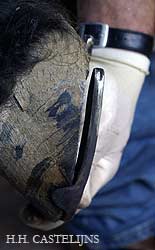
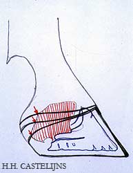
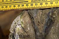
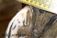
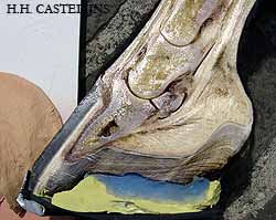
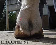
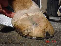
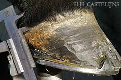
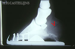
In pathological hooves, affected by D.P. displacement in the distal sense (sinkers) relative to the hoof capsule, due to laminitis, it is common to observe lateral and medial Q.C. on the same hoof. On palpation and measurement it is not uncommon to find the proximal border of the U.C. at or below the coronary band. In the case of sheared heels, the upwardly shunted heel is often affected by recurrent spontaneous Q.C.. When these are present, palpation of the U.C. and moving the U.C. outwards (abaxially) by hooking a finger axially to it, tends to elicit pain and opening of the proximal margins of the crack. (Fig. 3) The painful reaction is usually not elicited on the contra-lateral side (to the sheared heel) by the same manipulation. The proximal margin of the Q.C. is always near the highest point of the vertical distortion of the coronary band when observed from the side. Measurements of the free U.C. margin above the Q.C. by means of metric callipers, which is always done in the clinical practice of the author, reveals that when spontaneous cracks are present this distance is 15mm or less (-2 to 15mm). (Fig. 4) |
||||||||||||||||||||||
|
||||||||||||||||||||||
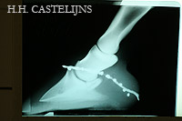
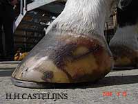
Sheared heels are usually due to angular deviations or, more frequently, rotational conformation defects of the limb such as inside rotations (toed in, medial sheared heel) and outside rotations (toed out, lateral sheared heel). A sheared heel is by definition longer than the contra-lateral heel, when measured from the coronary band to the ground or shoe, but absolute heel length (both normal and sheared) depend also on the palmar angle of the D.P. The palmar (plantar) angle of the D.P. is the angle of the distal margin of the D.P. with the ground as measured on a lateral medial radiograph with the focus of the X-Ray beam at ± 2,5cm above the distal border of the hoof. [Redden, 2003]. Palmar angle is determined by the tension of the deep digital flexor tendon, and can vary between front feet of the same horse, variations which occur during the development of the foal. [Van Heel, 2006] A larger palmar (plantar) angle goes together with longer heels and a more horizontal coronary band, as seen from the side, a foot with a smaller palmar (plantar) angle will present shorter heels and a more sloping coronary band. [Castelijns, 1998]. Free margin of the U.C. can be diminished in both types of feet due to upward displacement of the hoof wall at a heel relative to the ipsilateral palmar (plantar) process and attached U.C. and is the important parameter to keep in mind when addressing Q.C. In practical terms even a relatively low heel can be shunted upwards, relative to the structures (D.P., U.C.) it contains, if the D.P. has a low palmar (plantar) angle, hereby leaving little free U.C. margin and predisposing for spontaneous Q.C. as can often be seen in trotters and thoroughbreds. (Fig. 5) |
||||||||||||||||||||||
|
||||||||||||||||||||||
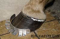
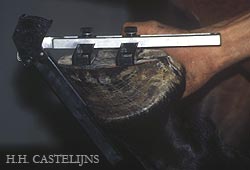
Manipulation of the coronary band at the quarters/heelsDorso-proximal distortion of the hoof wall at the quarter/heels can be corrected by full wall depth sub-coronary grooving, a technique already described by Laurino in 1932 for the realignment of the hoof capsule in the case of founder. [Pires, 1991] Sub-coronary grooving at the heels, although effective, is not very compatible with a heavy work load, can lead to excessive granulation tissue prolapsing outward, and does generally not rank high in customer satisfaction. Re-establishing a larger free margin of the U.C. at the site of a sheared heel can be done, and has been done, through the years by simpler means, Van Snow proposed it with a 24 hour soak on the unshod foot standing on a frog support. [Van Snow, 1991] The author uses a double trimming method inspired by Anz. [Anz, 2004] The hoof is first trimmed for the phalangeal alignment in the sagittal, frontal and horizontal planes. Flares and dishes of the hoof capsule (true distortion) are, as far as possible removed, the heels are trimmed back to the widest part of the frog. In the case of severe limb conformation defects (and therefore severe doubts) a Finnegans hoof gauge and/or a radiological protocol is used according to Caudron. [Caudron, 1998] (Fig. 6) |
||||||||||||||||||||||
|
||||||||||||||||||||||
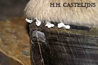
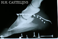
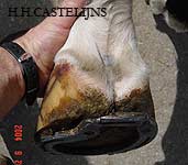
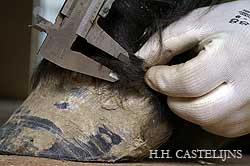
The shoe is then fit to shape and set hot. Before nailing on, a second trim is performed under the upwardly shunted quarter heel, which goes from 0mm at the ipsilateral toe (e.g. Inside toe for medial sheared heel) to an average of 7mm at the affected heel. The amount of heel which can be taken off in the second trim depends on the sole depth at the seat of corn, and on the severity of the upward displacement of the coronary band at the sheared heel. Usually enough sole depth is available which corresponds to the observations of Savoldi. [Savoldi, 2006] When not enough sole depth is available at the site of the affected heel for the second trim, the rest of the hoof wall can be raised with a rim pad or with a closed pad and filling material. Lack of sole depth at the heel to be lowered is more frequent in horses with a low or negative palmar (plantar) angle of the D.P. When nailing on the shoe and clinching it, the affected heel will lower itself on the shoe within minutes, making the original space, between the wall and the upper surface of the shoe, created by the second trim, disappear. The extent of the second trim at the heel will determine the increase of free margin of the U.C. above the affected hoof wall. This can be observed visually, palpated and measured with callipers. (Fig. 7. a, b, c, d, e) |
||||||||||||||||||||||
|
|
|||||||||||||||||||||
Fig. 7 a), b), c), d), e) - The double trim method permits the lowering of a single sheared heel, thereby obtaining a larger free margin of the ungular cartilage |
||||||||||||||||||||||
The amount of instant lowering that can be obtained in this way varies according to the size of the foot, the extent of the original vertical distortion of the sheared heel and the palmar angle of the D.P. (Fig. 8) |
||||||||||||||||||||||
|
||||||||||||||||||||||
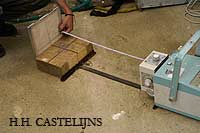
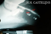
The author has measured a minimum of 3mm in small (28cm distal hoof wall circumference), thoroughbred type feet with a 1ºpalmar angle and moderate vertical distortion to 12mm in a larger (34cm circumference) with a 5º palmar angle and 20mm difference in height between the heels before treatment. If the height of a single heel/quarter can be lowered so easily, relative to the D.P., the ipsilateral U.C. and the contra-lateral heel, the question arises: can heels be deformed upward by raised heel shoeing or wedge pads? The question is of some importance as raised heel shoeing and wedge pads have been recommended for instance as therapeutic shoeing techniques for navicular syndrome. The reasoning being that by slightly flexing the distal inter-phalangeal joint, the angle the deep digital flexor forms around the navicular bursa and bone is less acute and therefore might relieve pressure and pain. [Denoix, 2005] If however these shoeing techniques deform the hoof capsule upwards relative to D.P. the biomechanical effect (flexing the joint) will be at least partially diminished. Results were measured by placing the D.P.s on the radiographs overlaying each other and measuring differences between the markers on the heels. (Fig.9) |
||||||||||||||||||||||
|
||||||||||||||||||||||
Besides heel wall displacement, the dorsal wall angle was measured with a Dallmer hoof-gauge; other data recorded where name, breed, age, sex, use, height and estimated weight and hoof size. The Dallmer hoof-gauge was used because of it’s reliability. [Moleman, 2002] Measuring the upward displacement on the overlaying radiographs was done where it was largest, measuring from the top of each line (or dot) to the top of the overlaying one. Results are reported in Table 1. |
||||||||||||||||||||||
Discussion:All limbs showed vertical displacement when placed on a 20mm high wedge. It should be noted that displacement was instantaneous (the time to place the foot on the wedge, pick up the other foot and shoot the x-ray). It might well be that over time, upward displacement increases, as it also might under increased load. Horses’ feet in Tuscany in June 2006 were relatively dry, (estimated shore values of the frogs upwards of 50A), it is reasonable to expect humid feet to show even more capacity for distortion. Placing a wedge under a single heel, with the same body weight pushing down on the limb will probably result in more vertical displacement of that particular heel. The heels of the horse’s foot have a relative large amount of flexibility in the proximal distal (vertical) sense. This can be explained by the anatomical features of the foot: a discontinuity of the hoof capsule between the heels with highly mobile structures interposed, such as the frog, digital cushion, venous and arterial plexa and fibro-cartilaginous connective tissue. In other words although the dorsal wall is intimately attached to the dorsal aspect of the D.P., the laminar suspension apparatus in the heel area is attached much more loosely. Functionally this arrangement serves the bare footed horse well as the hoof capsule at the heels can adapt itself to the uneven footing. When trimming or shoeing modifications in the sagitale plane of the foot (toe to heels) are contemplated, it is important to keep this vertical mobility, and capacity for vertical shunting of the heels, in mind. Leaving heels (too) long or raising them with raised heel shoes does not change the palmar/plantar angle of the D.P. to the same degree because of immediate upward displacement of the heels. It also stimulates a greater wear of the heels on the upper surface of the shoe so that the resulting change in the flexion/extension of the distal inter-phalangeal joint is probably even more reduced in the course of the shoeing interval. Recruiting other palmar/plantar structures of the hoof into weight bearing might give better results as presumably this will unload the heels at least partially. This can be done with special pads and polymer filling [Renieri,2002] [Castelijns, 2005] (Fig. 10) |
||||||||||||||||||||||
|
||||||||||||||||||||||
Preventive and therapeutic shoeing for podotrocleosis can always benefit from ease of break over with for example rolled, rockered, set back and open toe shoes.[Denoix,2005] In the prevention and treatment of spontaneous Quarter/Heel cracks it is important to lower the coronary band at the sheared side. The object is to get more than 15mm of free U.C. above the affected side (on average 30-32cm circumference hoof). As most feet with a sheared heel belong to a limb with a conformation defect, (rotational angular), these feet have a tendency to deform their atrophied heel upward again, and the double trim method usually has to be applied to some degree at each shoeing. Shoeing intervals should also be kept reasonably short (5-6 weeks, compatible with hoof growth). Spontaneous Q.C. cause pain and often lameness because they develop from the inside out and their deeper margins may pinch the underlying laminar tissue or can even become infected. Debridement, cleaning and fixation are therefore important in the more extended Q.C.. Modern hoof repair materials are of great help in these procedures, while care should be given to place drainage in the deeper parts of freshly debrided Q.C. [Magri, 1998, Duvernay,2000] Correction of the cause; limitation of the outward U.C. expansion by the hoof wall, has to be addressed if recurences are to be avoided, especially in full working conditions of the modern sport horse. * Prontobario esofago, Bracco s.p.a. – Via E. Folli, 50 Milano The author would like to thank Michael T. Savoldi for his images and Simone Bizzi, C.F. for his physical assistance. |
||||||||||||||||||||||
Bibliography:
|
||||||||||||||||||||||
 7A
7A 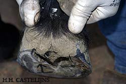 7C
7C 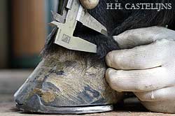 7E
7E