Foal development: Angular and rotational deformities around the fetlock.
|
I Introduction:
Flexural, angular and rotational deformities may be present at birth but take several months of treatment to correct.
The first six months of the foal’s life are a time of rapid development, during which 60% of the increase in height after birth takes place; in these months deformities (especially flexural), are often acquired as part of the general syndrome of developmental orthopaedic disease (DOD), which also includes physitis and osteochondrosis.
Breeders expect to produce equine athletes, or at least yearlings which will be considered potentially so by buyers at the sales; “correct” leg conformation is important to obtain a good price at the sales, sustain a successful training regime in the young adult, to attain a high score at the shows, and perhaps most important, to prevent lameness during a long career of the sport or racehorse.
Leg conformation, and it’s defects, is determined before birth by genetics of the parents.
The mare’s influence is of course both genetic, although it usually takes several offspring to notice, as environmental, therefore management of the mare is an important factor.
Obese mares (during pregnancy), as seems to be the rule with a lot of P.R.E. (Andalusian) breeders in Spain, tend to have a higher incidence of foals with angular deviations at birth. The higher incidence, specifically at the level of the carpal and tarsal joints, with a lot of “windswept” foals (one hock varus the opposite valgus), could be due to the reduced uterine space caused by excessive abdominal fat.
|
II . Evaluation of limb conformation.
A. Evaluation of the foal for it’s conformation at, or soon after, birth is useful in all foals and essential in high risk cases: twins, premature, dystocias, and previous history.
B. Evaluation should take into account:
- That a foal is not an adult:
a. A base wide, outwardly rotated stance of the front legs is perfectly normal in lighter (dolicomorphous) breeds, as it increases stability of these long legged narrow breasted babies ¹ , and tends to “correct” itself with time.
b. This stance should not be confused with a valgus fetlock when seen from the front. In fact the best view for catching angular, and especially rotational deviations, of the front limbs is the skyline view, looking down from the shoulder to the toe, evaluating how the radius aligns with the cannon bone and the cannon bone with the pastern and the hoof.
- The age of the foal.
a. Growth plate closure times can be defined functionally, radiologically and hystologically. For correction purposes the first are essential.
b. Distal growth plates (proximal of PII, proximal of PI, distal of MC/MTIII) close functionally within the first 3 months, followed by the distal GP of the tibia and finally the distal GP of the radius ²
c. Food intake changes naturally with age; D.O.D. is due mostly to excess, not only of protein but also of energy (carbohydrates). Limiting energy intake takes a different approach in the new born as opposed to a 3 month old, which is grazing on lush spring pastures and/or might have access to crib feeding.
- Body condition score and weight.
An upright pastern is normal in the light weight newborn, the fetlock should naturally extend further (drop) with increase in body weight.
- The breed.
For example:
Quarter horses and Andalusians are already “stocky” at birth (bracimorphous) and have higher incidence of varus deviations than, for instance, thoroughbred foals.
A slight inward rotation of the lower hind leg is as lot more serious in a Standard bred trotter than in other breeds as it will probably lead to a cross gaited movement and interference at lower to medium speeds in future training.
C. Evaluation should be performed viewing the foal/limb:
- From the side to spot abnormalities in the sagittal plane (flexural deformities).
- From the front or the back to spot angular deviations in the frontal plane.
- From above to look for axial rotations in the horizontal plane.
- Bear in mind that the same foal, indeed the same limb, often presents a combination of flexural, angular and rotational deformities.
|
|
Table I.
Growth plate activity and closure times in months. ²
|
growth plate: |
End of fast growth |
Radiological closure |
Distal tibia |
8 |
17-24 |
Distal radius |
8-10 |
20-42 |
Distal MC/MTIII |
3-4 |
6-15 |
Proximal PI |
2-3 |
12-15 |
Proximal PII |
2-3 |
8-12 |
|
III Treatment of angular and rotational deformations.
A. Angular deviations (in the frontal plane) are defined as:
- Valgus whereby the segment of the limb, immediately distal to the affected joint/growth plate, is deviated laterally.
- Varus ; whereby the limb segment below the deviation is deviated medially.
- Bear in mind that the same limb may present valgus and varus deviations at different points: e.g. carpus valgus with fetlock varus.
B. Varus and valgus deviations can be observed by:
- Viewing the limb from the front or the hind (often useful for evaluating fore limbs too).
- Viewing the limbs from above, positioning oneself close to the shoulder or the hip of the foal.
- On the fore limbs, and to judge cannon bone – pastern alignment, by picking up the fore limb at the distal end of the cannon bone, whilst letting the digit hang down freely.
- A long ruler or a farrier’s rasp can be useful as a visual guide, when in doubt.
- Dorso-palmar/plantar x-rays permit accurate measurements, the assessment of ossification of carpal bones and the exact area where deviation takes place; the use of long cassettes is recommended. (Assessment of ossification of tarsal bones is best done with latero medial x-rays of the hock.)
NOTE: Beware of assessing a fetlock as valgus, on an outwardly rotated fore limb. For there to be a valgus fetlock, the pastern has to be truly deviated laterally relative to the cannon bone.
C. Varus deviations tend to get worse with growth, when left untreated, valgus deviations tend to self correct with widening of the upper body.
D. Treatment of angular deviations depends on active growth plates and includes:
1. Trimming:
- i. Correct hoof flares on the side of the hoof to which the lower limb deviates to.
- ii. Lower the hoof on the side the distal limb deviates to, only if this side is too high.
- iii. Trim the frog, which is comparatively larger in a foal than in an adult horse, asymmetrically – more on the side the limb deviates to.
- iv. Thin the wall on the side of the deviation.
- v. Ideally it is better to trim a little often, than a lot at long intervals.
- vi. With short hooves and/or larger deviations, there is a limit to what you can achieve with trimming.
2. Lateral or medial extensions to the hooves.
With polymers like polyurethanes (e.g. superfast, adhere) or polymethylmetacrylates (PMMA) (Equilox, Bond n’Flex, TopGum etc.)
- i. Polyurethanes like Superfast are quick setting and wear resistant.
- ii. PMMAs have excellent adhesion, but should be reinforced with glass – carbon or Kevlar fibres, to increase their resistance to wear.
- iii. With foal shoes:
- a. Foal cuff type shoes on the market include Mustad Baby Glu (cyanolitic glue), Dallmer (Dallric in the U.S.A.) foal shoes (epoxy glue), and Ibex foal shoes (cyanolitic glue).
- b. All these foal shoes can also be applied with polyurethane adherents (superfast, adhere from Vettec) or Polymethylmetacrylates (PMMA’s like Equilox, Bond N’ Flex, Top Gum etc.)
- c. All glues except cyanolitic ones, release heat, care should be taken to protect the hairline with duct tape.
- d. The hoof’s surface needs to be mechanically cleaned (sand paper) and dried (hairdryer) if wet.
- e. All cuff type shoes should be removed within 7-8 days after application on the newborn or days old foal, as they will not allow for hoof expansion because foal hooves grow twice as fast as adult hooves and are narrower at the bottom then at the coronary band in the newborn.
- f. A useful tip is to write the application, or the recommended removal, date on the shoe (see table II).
|
Table II .
Foal shoe removal times relative to the foal’s age on application |
Age (days) |
Remove within (days) |
1 |
7 |
7 |
10 |
20 |
14 |
45 |
20 |
|
3. Surgery.
- i. Periostal stripping on the “hollow” side, at the growth plate where the deviation takes place, that is, on the side which has an angle of less than 180°, the procedure is done under general anaesthesia.
- ii. Transcutaneous growth plate stimulation with an 18G, 40mm needle. This can be done with local anaesthesia, surgical prep of the overlaying skin and approximately 10mm deep stabs into the growth plate, on the side where growth needs to be stimulated (concave side), in a fan shaped pattern with as the apex the single cutaneous perforation and underlying periostum. 3 Advantages of this technique relative to periostal stripping are that it can be done at the farm, and doesn’t cause as much periostal reaction or scars.
- iii. Transphyseal bridging with screws and wire, screws and plates. (General anaesthesia).
- iv. Periostal stripping, or transcutaneous stimulation of the growth plate, aims at increasing growth on the ipsilateral side, does not over correct, is repeatable after a 6 week interval, and depends on an active growth phase of the treated physis.
- v. Transphyseal bridging is a growth retardation procedure on the side with a larger angle than 180°, the implants have to be removed (2 nd surgery) as they may overcorrect, they still work at slower (later) phases of physeal growth.
4. Combination of surgery and hoof extensions.
- i. Periostal stripping or transcutaneous growth plate stimulation work for 4 to 6 weeks and are synergetic with hoof extensions on the opposite side of the limb, e.g. lateral stripping of the distal physis of the radius and medial hoof extension for the correction of severe carpus valgus.
E. A rotational deviation (in the horizontal plane) is defined as an axial rotation of a limb segment relative to the segment proximal to it.
- It can be inward or outward relative to; e.g. inward rotation of the digit relative to the cannon bone.
- Rotations can best be observed from above, looking downward along the limb.
- Rotations of the digit, relative to the cannon bone, can be judged by picking up the limb loosely at the distal end of the cannon bone, and letting the digit hang.
- i. Observe the direction of the frog, relative to a line perpendicular to the ground.
- ii. Rotate the hoof capsule gently, it will rotate more, and with greater ease, in the direction of the rotation of the digit.
F. Treatment of rotational deviations.
- Only rotations of the digit relative to the cannon bone can be corrected by trimming or hoof extensions.
- Trimming or hoof extensions should extend the toe-quarter area of the hoof, opposite to the direction of the lower limbs rotation; e.g. a lateral toe extension, to correct an inward rotation of the digit relative to the cannon bone.
- Outward rotations of the entire fore limbs from the elbow down, present at birth and in the first weeks – months of life, tend to self correct with growth and widening of the thorax, and should therefore not be treated with hoof modifications in the first 6 months.
- Inward rotations tend to get worse with growth, and should be treated more aggressively than outward rotations, which have a tendency to self correct.
|
IV: Treatment of angular and rotational deviations of the fetlock.
NOTE: Lower limb growth plates, around the fetlock, close functionally a lot earlier than the distal plates of the tibia and radius; priority should therefore be given to get the lower limbs well aligned at the fetlock within the first 3-4 months of the foal’s life. (See table I).
|
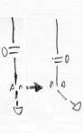
A. Fetlock varus. |
 |
|
1. Frequent condition in all breeds, including trotters, warmbloods, Andalusians and heavy breeds but also of the hinds of thoroughbreds.
2. Can be masked, upon superficial evaluation, by valgus carpus or hock, or by outside rotation of the entire limb.
3. Needs conscientious treatment within the first three months, because of lower growth plates closure times. Tends to worsen with growth.
4. Can be present at birth (check for latero medial laxity at the fetlock).
5. Often acquired during growth in heavy, wide breasted foals or as a consequence of weight bearing on the opposite limb to an injured limb.
6. Trim medially, save hoof laterally.
7. Extend with a polymer extension to the lateral side.
- a. Ideally to the extent to where the outside of a hoof, belonging to a normal digit, would reach.
- b. Extend the whole side wall, including the heels.
- c. Beware of pinching or burning the coronary band at the heels with the fast setting and heat producing polymers.
- d. Do not let it grow forward too much, as it will then become a lateral toe extension, favouring outward rotation of the digit. Trim regularly.
8. Extend with a lateral extension foal shoe.
- a. With large degree of varus (>3-4°), present at birth, or acquired from overload on one limb, apply lateral extension cuff type foal shoes, as these can usually extend further than extensions out of pure polymer, they also “pull on” the whole hoof, not only the side wall.
- b. Wide lateral extensions with foal shoes, which can have as much ground surface as the hoof itself, are also called for when the fetlock varus belongs to a limb which has an added varus at the carpus or hock. e.g. “Windswept” foals which are born with one hock varus and the opposite valgus, often have a varus fetlock on their varus hocked limb, both joints react well to a wide lateral extension, applied in the first days after birth.
- c. When using cuff type shoes, set the cuff a bit wide on the side of the extension, as the hoof wall there tends to be a little straighter.
- d. Respect foal shoe removal times (table II).
- e. Do not raise and then extend the lateral wall, but only extend it.
9. Surgery
- a. Stripping or transcutaneaous stimulation of the medial aspect of the distal growth plate of the cannon bone and/or the proximal G.P. of the first phalanx.
- i. Take dorso palmar x-rays to decide, and as a baseline to judge improvement.
- ii. In the author’s experience, periostal stripping of these growth plates results in more periostal reaction and visible swelling than stripping of the higher growth plates.
- iii. To be done in the first three months of life.
- b.Transphyseal bridging of the lateral aspect of the distal G.P. of the cannon bone, can be attempted for severe deviations (> 4-5°), and when time is running out (10-14 weeks of age).
|
|
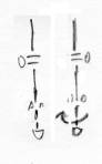
- True fetlock valgus is a rare condition.
- Check by hanging the limb.
- Tendency to self correct permits less aggressive treatment.
- Check for latero medial laxity at the fetlock in the newborn.
- Trim laterally, save medially.
- Small medial extensions with polymers may be of help.
i.
Do not have extension on the medial wall in the toe area, as this easily causes inward rotation of the digit.
- Surgery is rarely called for
i.
Lateral stripping or physeal stimulation of distal GP of MC/MT III and/or of proximal GP of PI.
ii.
Medial transphyseal bridging of distal GP of MC/MT III
- Do not over correct!
|
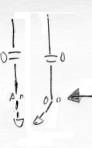
C. Inward rotation of the digit, relative to the cannon bone. |
|
|
- Frequent condition, often acquired during (fast) growth.
- Contributing factors may be:
i. Stocky heavy breeds or individuals.
ii. Hilly paddocks.
iii. DIP hypo extension (club foot): most club feet are also turned (rotated) inward. iv. Mistaken “correction” of outwardly rotated, base wide stance of entire limb.
- Trim medial toe, sometimes reduce lateral heel flare, save or leave extra hoof at the lateral toe area.
- When not enough hoof is available, extend lateral toe with polymers.
- Keep monitoring the condition as it tends to grow worse with growth and increase in width/weight.
|
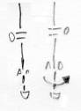
D. Outward rotation of the digit, relative to the cannon bone. |
 |
|
- Rare condition on the fore limbs, more frequent on hind limbs.
- Check your diagnosis by hanging the limb, looking for the way the frog points and manipulating the hoof outwards and inwards.
- Tendency to self correct, treat less aggressively.
- Trim lateral toe, reduce flare on medial heel if present, save medial toe.
- Extend medial toe slightly with a polymer if the hoof is worn too much in this area.
- Monitor, do not over correct.
|
References
- Curtis, S.: From Foal to Racehorse. Newmarket Farriery Consultancy.Newmarket,1999.
- Betsch, J.M.: Reconnaissance, évaluation et gestion des déviations angulaires du poulain. Pratique vétérinaire équine, volume 37, 45-59,2005
- Colles, C.M.: Physeal Stimulation for the Correction of Angular Limb Deformities, Proceedings. Beva congress 2006 page 319. Kildare Ireland
- Auer, J.A.: Angular limb deformities, Flexural limb deformities. Chapters 89-90, pp 1130-1165, Equine Surgery III d edition, Saunders Elsevier, St. Louis MO, 2006.
|
|