Trimming and Shoeing in the Frontal and Horizontal PlanesAbbreviations:
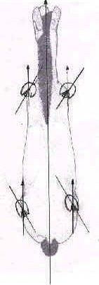
1. IntroductionAll three-dimensional objects or organisms can be evaluated in three planes: sagittal (toe to heels or dorso/palmar/plantar for the hoof), frontal (F.P.) (latero-medial (L.M.)) and horizontal (H.P.) (diagonally across the hoof). |
||||
|
||||
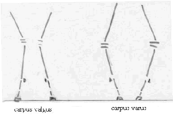
This paper seeks to address the relationship between hoof shape and (leg) conformation in the F.P. and H.P.; and the consequences of trimming and shoeing in these planes. 2. Conformation in the Frontal and Horizontal PlanesThe ideal conformation of a horse may vary by breed and by observer but to a large extent there is agreement on what constitute conformation faults: Angular Deviations (A.D.) in the F.P. and Rotational Deviations (R.D.) in the H.P. The first can best be evaluated by viewing the horse from the front or the back, the second by looking down the limb standing close to the horse. A.D.s are defined as valgus when the segment distal to the A.D. is deviated laterally and varus when the distal segment in deviated medially, preceded by the joint site of the A.D. |
||||
|
||||
R.D.s take the name of the segment, which is rotated either inwards or outwards. |
||||
|
||||
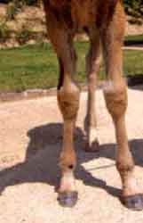
Both type of deviations cause joints to work at abnormal angles to the sagittal plane of the horse, which is also the plane in which the horse moves when not turning, equine limb joints having evolved to function mainly as hinges at 90° to the sagittal plane. The possibility of correcting conformation faults is dependent on having active growth and is attempted by influencing growth plate development and therefore should be attempted at an early age (first year of life). Correction of conformation defects is not possible and should not be attempted in the adult horse. |
||||
|
||||
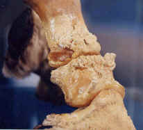
3. Consequences of A.D.s and R.D.sA.D.s cause the affected joints to be loaded unevenly in the L.M. sense, with both increased loading on the articular cartilages on the concave side and increased tension on the collateral ligaments on the convex side. A horse affected with R.D.s, when moving in a straight line, puts an analogous amount of stress on the affected joints as a straight horse who moves in a circle. |
||||
|
||||
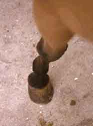
Another consequence of these conformation defects is that they tend to alter the flight path of the limb during suspension. For example; outward rotations tend to favour a medial deviation from the sagittal plane of the lower leg, when swinging past the opposite limb, while inward rotations may cause an outward swing, typically landing on the medial heel and breaking over on the outside toe of the hoof. |
||||
|
||||
Most experienced farriers and veterinarians will agree with the observation that horses with these types of conformation defects tend to show a high incidence of articular pathology, including capsulitis, entheseophyte formations, ringbone, navicular syndrome (note that epidemiologically it is mostly horses who perform a lot of turns who have a high incidence of navicular disease). Other common pathologies include splints and bone spavin, while the hoof often suffers from quarter cracks. |
||||
|
||||
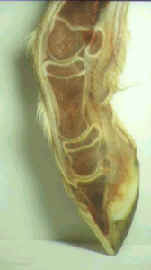
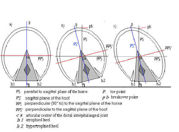
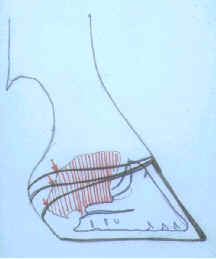
4. Hoof shape in relation to conformationThe hoof capsule responds in a predictable way to the particular conformation of the limb it is attached to. In the case of a pure A.D. the hoof capsule is relatively atrophied on the opposite side of the deviation, e.g. a lateral deviation tends to cause a hoof with relative medial atrophy. There is a functional advantage to the affected horse in this adaptation of the hoof capsule. By underdeveloping the side opposite to the direction of the deviation, the joint spaces tend to remain parallel, with more evenly spread L.M. pressure across their surface and with more even tension on the collateral ligaments during weight bearing. |
||||
|
||||
In the case of R.D.s, the hoof capsule adapts by atrophying diagonally across the hoof, along a line that is parallel to the sagittal plane of the horse, e.g. an inward rotation causes medial heel and outside toe atrophy and when those relatively atrophied hoof parts are connected by a line this line is parallel to the sagittal plane of the horse, but not to the sagittal plane of the hoof. Again there is a functional, biomechanical, advantage to the affected horse in this arrangement. By shortening the distance between both the atrophied heel and the atrophied toe to the centre of the distal interphalangeal joint, a joint that does not square up with the horse's body, the torque on this joint is diminished both in the heel landing and the breakover phase. |
||||
|
||||
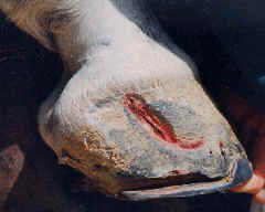
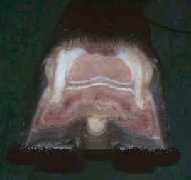
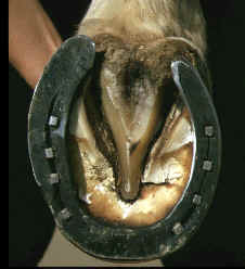
5. Trimming and shoeing the limb with conformation defectsGood farriery aims at minimising excessive joint stress in horses with A.D.s and R.D.s. L.M. trimming is carried out with the objective of keeping joints evenly spaced. In order to achieve this it is usually appropriate to recognise and respect the hoof shape, even though in severe deviations this shape looks out of the ordinary. With some experience it becomes possible to correlate the "abnormalities" in the hoof shape to the conformation of the limb it is attached to. Shoeing to fit the hoof and not the other way round is, of course, a very old, “golden” rule in farriery. Respecting the adaptation of the hoof shape to conformation can be done by looking at the white line, which will indicate "true" hoof shape better then the outside wall. The outside wall in horses with A.D.s and R.D.s often deforms easily with the hypertrophied parts flaring outwards, particularly when too much time passes between two shoeings/trims. These deformations (flares) are characterised by outwardly bent hoof tubules (concave hoof wall) and should be corrected by rasping the distal wall to obtain a straight proximo-distal profile. |
||||
|
||||
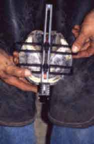
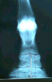
L.M. trimming the bottom of the foot at 90° to the central hoof tubule with the help of a Finnegan's hoof gauge has been shown to result in evenly spaced distal joints in the equine limb by checking with a radiological protocol, which of course remains the most precise means of establishing appropriate L.M. balance in difficult cases. |
||||
|
||||
Since relatively atrophied hoof parts expand less and are usually thinner, these should preferably not be burdened with clips or nails, which are better concentrated at the overdeveloped parts of the hoof capsule. Compensating with extra width (extensions) of the shoe at the site of the hoof's underdeveloped parts should also be approached with caution. Although it makes for a more symmetrical hoof and shoe outline, setting a shoe wide and/or long, particularly under an atrophied heel may cause the moment arm to the centre of the distal interphalangeal joint to increase and may actually cause inward and upward pressure on the hoof wall at the affected heel. These heels, which are easily shunted upwards and sheared, are often the sites of spontaneous hoof cracks, due to ungular cartilage meeting the upwardly shunted coronary band from the inside, when bending outwards during weight bearing. |
||||
|
||||
Horseshoes that bring L.M. breakover closer to the hoof's centre will diminish joint stress and should perhaps be used preventively in horses with conformation defects and not only after arthrosis has set in. Finally, as mentioned before, no matter how perfectly adapted a shoeing job is on the day of shoeing, if too much time passes and too much hoof growth takes place before the next shoeing, the hoofs of horses with conformation defects in the F.P. and the H.P. will easily become distorted with flares and the long biomechanical levers will jar and stretch joints and ligaments. 6. Appropriate use of lateral or medial extensionsLateral or medial extensions, although not recommended for “corrections” of conformation defects of adult horses, do have appropriate uses. They include the correction of foals, influencing gaits (e.g. in standardbreds) and as therapy for certain pathologies. Collateral ligament injuries and injuries to one branch of the suspensory ligament may benefit from an extension on the side of the injury. Bony lesions with clear medial or lateral location, e.g. medial bone spavin, might improve with the extension placed on the opposite side of the lesion. The therapeutic action is due to the fact that on soft arena-type surfaces the side with the wide web (extension) will not sink as deep into the ground as the narrow branch of the shoe, therefore diminishing ligament strain on the extension side and preventing articular pinching on the opposite side. |
||||
|
||||
7. ConclusionWhen holding an asymmetrical hoof, it is good practice to put it down and take the time to analyse the conformation of the limb it is attached to. Farriers and veterinarians should beware of the urge to always obtain nice, symmetrical, "normal" feet, which might be more pleasing to the eye with the horse standing, but that might not respond to the biomechanical needs of a horse affected by angular or rotational deviations. |
||||
|
||||
*Bibliography on request. |

Hans Castelijns
D.V.M - Certified Farrier