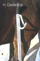
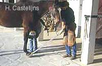
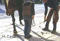
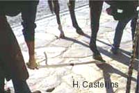
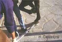
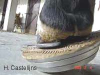
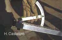
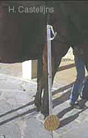
A Digital Extension Device for extension test in lameness examinationsThe Dorsal digital extension test is considered useful by many clinicians when doing a lameness examination of the front limbs. Traditionally the limb under examination is placed on the extremity of a long board, an assistant picks up the controlateral limb and the opposite end of the board is slowly raised extending the digital joints, increasing the tension on the deep digital flexor tendon (DDFT), its carpal or inferior check ligament, the palmar angle of the DDFT around the navicular bursa and distal sesamoid (D.S.), and the collateral and impart ligaments of the D.S.. Apart from practical problems like bending or breaking boards, pinched fingers, and slipping of the subject's foot, evaluation of positive or negative responses are only standardized, if at all, with a personal protocol, e.g. by using always the same board, careful placing of the foot at its extremity and deciding on a height to be reached at the opposite end (e.g. The examiner's knee) [Debrosse]. Elevating the foot sideways, either laterally or medially, with the use of a board, further on improperly discussed as lateral and medial extension, is considered by some clinicians useful in the diagnosis of arthrosis - arthritis in the digital joints, painful collateral ligaments of these joints, sources of subcondral pain with a lateral or medial location in the joints and probably of lesions of the distal part of the DDFT which as has been recently shown with magnetic resonance imaging [Dyson] and sophisticated ultrasound studies [Denoit] might more often be the source of "Navicular syndrome" or even less specified "palmar hoof pain" then previously thought, bearing in mind that in its very distal portion the DDFT, with its characteristic -8 - shape, is quite wide with a definite lateral and medial component.It is also interesting to note that lateral or medial elevation of the foot does not only cause movement, pinchming and tensions in the frontal plane (side to side) of the components of the proximal and especially the distal interfalangeal Joints (PIP an DIP) but also in the horizontal plane with a strong rotational movement of the distal phalanx (PIII) relative to the intermediate phalanx (PII) and the D.S.; this movement has been elegantly studied in vitro and described with the name of colateromotion [Denoit]. From the above it could be concluded that digital extension test are certainly not very specific (as to the exact site of pain or lesion) and until now not even very sensitive although this certainly varies with the experience of the clinician. This does not necessarily diminish the practical value of digital extension tests, especially in regard to some treatment options as for instance therapeutic farriery. It is for example likely that a horse with an intolerance to dorsal extension of the interphalangeal joints will benefit from specific farriery techniques such as bringing back dorsal break over, egg bar shoes and reverse shoeing which limit digital extension in deep ground and or raised heels. [Castelijns].Increasing the ease of use and specially the objectivity of the digital extension test by making it quantifiable has now been made possible with a Digital Extension on Device [DED] manifactured by an Italian company [Colleoni] which also manifactures high quality Aluminium horse shoes. The DED consists of three round plates of which the middle one attaches to a 4x4x120 cm long handle which at its opposite extremity has a rubber covered grip and close to this a protractor with a needle which incorporates a spirit level. The top plate is covered with rubber to prevent slipping and has a diameter of 21 cm; a set of internal ball bearings permits the middle plate (and its attached handle) to rotate relative to the top and bottom plates making it possible to do Dorsal Lateral and Medial extensions without having to replace the foot on the DED. The sides of the plates are bevelled at a 45° angle, reducing the diameter of the ground surface of the bottom plate to 16,5 cm, making lifting easier for the clinician.When reaching the maximum degree of extension the horse tolerates (dorsally), or when the opposite side of the foot is starting to lift off the plate (lateral and medial extension), the needle is placed water level and stays there thanks to a friction device, enabling reading of the measurement after the handle is lowered again. After reading the needle position on the protractor (with a c.c. milled scale of 0 - 60°), the handle can be swivelled to the next extension and the next measurement taken. |
|
Hans Castelijns
D.V.M - Certified Farrier