Hoof Deformation in the Vertical Sense: Causes, Consequences and ManagementIntroduction:The hoof, as is well known, permits the heels to open and close on the horizontal plane in correspondence with each stance and swing phase of the limb.
|
||||
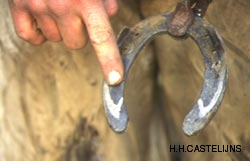
Apart from the horizontal hoof mechanism, the hoof also shows ample movements of the heels in the vertical direction relative to the internal structures of the foot, and of each heel relative to it’s opposite.
In fact the vertical movements that can be created at the heels by manipulating an unshod hoof are larger than the opening and closing movements of the latter. In the case of horses shod with traditional shoes, the vertical movements of the heels are limited by the rigidity of the shoes, leaving only the horizontal hoof mechanism. |
||||
|
||||
Vertical deformation of the hoof capsule:Horses with conformation defects, such as angular (valgus, varus), and rotational deviations (internal - “toes in”, external - “toes out”) and with combinations of these defects, invariably have asymmetrical hoofs. There is indeed a direct correlation between the shape of the hoof and the conformation of the limb it belongs to. |
||||
|
||||
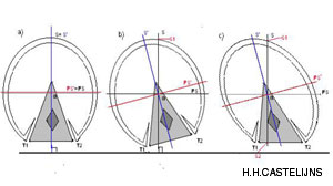
The adjustments of the bottom of the hoof are useful for the horse because they reduce the biomechanical levers on the distal joints; “hinge” joints basically evolved for flexion and extension movements in the horse’s sagittal plane. (Castelijns, 1994) |
||||
|
||||
The objective of correct trimming is to keep interphalangeal joints spaced evenly. (Serteyn, 1995) |
||||
|
||||
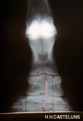
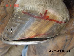
Apart from the useful adjustment of the shape of the hoof to the defective stance, these hooves easily become deformed. The deformation of the hoof capsule, or parts of it, can be recognised by observing the course of the horn tubules; if the latter are bent between the coronet and the distal edge of the hoof, this means there is real deformation which should not be mistaken for the physiological adaptation of the hoof to the limb’s stance. Examples are: distal walls with a concave profile, under-run heels and flaring of the sidewalls.
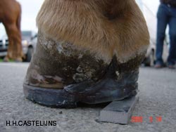
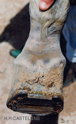
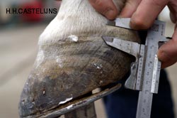
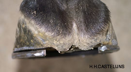
The hoof capsule also is easily deformed in a vertical direction, especially in its posterior areas where the laminar mechanism is not anchored to the rigid back of the third phalanx, but to the flexible structures of the ungular cartilages. The vertical deformation of the heels can be noticed by observing, and palpating, the coronet and its course (seen from the side), comparing the respective heights of the two bulbs and by measuring the length of each heel from the coronet to the distal edge of the hoof. |
||||
|
||||
Consequences:The consequences of a vertically displaced heel/bulb, a situation which is usually accompanied by a locally straight (hypo-conical) wall, may be the following:
In practice the consequences, described above in order of growing severity are primary caused by one or more conformation defects of the limb in question, and are made worse by excessive length of the hoof (the longer it is, the more subject to deformation it becomes, both in a transversal and vertical direction). |
||||
|
||||
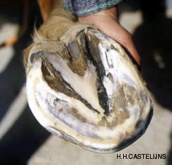
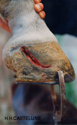
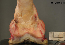
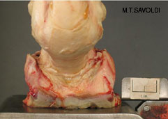
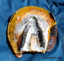
In a symmetric, normally conic foot, with coronets that follow a linear course (seen from the side), there is ample room for expansion of the ungular cartilages above the proximal edge of the hoof. If the wall is strongly deformed in a vertical and proximal direction (upwards), this space is no longer there; the inevitable expansion of the ungular cartilage will traumatise the coronary corion and the proximal laminae from the inside out, causing an often painful and bleeding quarter-crack. |
||||
|
||||
The clinical examination of the foot affected by a spontaneous quarter crack, easily reveals that:
|
||||
|
||||
Management of vertically displaced heels:The marked asymmetry of certain hooves, and the ease with which as a consequence one of the heels verticalizes and shunts upwards in these hooves, is due to conformation defects of the limb the hoof belongs to. |
||||
|
||||
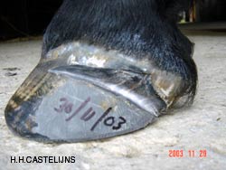
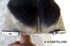
In spite of its effectiveness, this method is relatively harsh and radical; therefore it is unlikely that it will be enthusiastically accepted by the owners unless it is absolutely essential. As an alternative it is possible to achieve the descent of an upwardly shunted bulb/heel of up to 7 mm on average by adopting a more conservative method. After trimming the foot (preferably hot), respecting the digital axis, a further trim is performed which goes from the toe to the ipsilateral heel below the vertically displaced coronary band (Anz, 2004). When placing the shoe on the foot, it will seat on only two thirds of the hoof wall, with a progressive space below the portion of the wall which needs to be lowered. When nailing, it is advisable to first use the nail holes of the portions of the shoe which are in contact with the hoof; that is, those on one side and the toe nail hole on the opposite side. By allowing the affected limb to rest on the ground, the time needed to shoe the other limb(s) will be enough to have up to +/- 7mm of the space between the hoof wall and the shoe disappear through the descent of the upwardly shunted wall. |
||||
|
||||
With this method it is possible to obtain rather satisfactory corrections of vertically deformed hoof walls by relatively simple means. For severe vertical displacements, it can be useful to recruit additional parts of the hoof, such as frog, sole and contro-lateral bar (to the upwardly shunted heel), into weight bearing. For instance this can be done with the use of straight or heart bar shoes, pads and sole support, initially always leaving a space between the shoe and the verticalized wall (Castelijns, 2005). |
||||
|
||||
Conclusions:Horses with serious conformation flaws of their limbs, (especially rotations and even more so if in combination with angular defects), have a tendency to suffer from hoof deformations in a vertical sense. Therefore the solutions described in this article may prove not to be permanent, but part of the continuous preventive management of the hooves and limbs, aimed towards extending the useful life of the horse. “The (spontaneous) quarter cracks are like old friends; when you least expect them they appear again.” Special Thanks to Michael T. Savoldi |
||||
Hans Castelijns
D.V.M - Certified Farrier